Bone of the Front The frontal bone is the solitary bone that contributes to the formation of the forehead. Between the brows, in its anterior midline, there is a tiny dip called the glabella (see Figure 3). The frontal bone also serves as the orbit's supraorbital border. The supraorbital foramen, located at the center of this border, is the passageway for a sensory nerve to the forehead. Just above each supraorbital boundary, the frontal bone thickens, generating rounded brow ridges. These are placed slightly below the brows and vary in size across people, however men often have bigger ones. The frontal bone extends posteriorly into the cranial cavity. This flattened area serves as both the roof and the floor of the orbit below and the anterior cerebral cavity above (see Figure 6b).
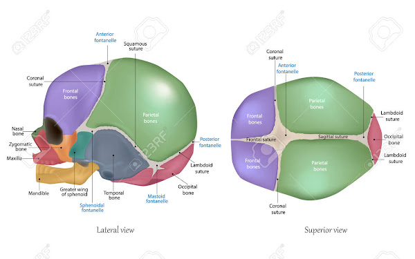
The cranial bones of a newborn child are fragile and underdeveloped. They contain a trace of calcium, are poorly defined, and feature six regions of partial ossification known as fontanels (Fig. 20-4). Two fontanels are located in the midsagittal plane, next to the superior and posterior parietal bones. The anterior fontanel is placed near the bregma at the confluence of two parietal bones and one frontal bone. The posterior fontanel is placed posteriorly and in the midsagittal plane, at the region designated lambda in Fig. 20-2. Additionally, each side has two fontanels at the inferior angles of the parietal bones. Each sphenoidal fontanel is located near the pterion, whereas the mastoid fontanels are located near the asteria. Normally, the posterior and sphenoidal fontanels close during the first and third months of infancy, whereas the anterior and mastoid fontanels shut during the second year of life. The skull grows fast in size and density over the first five or six years, then gradually increases until it reaches adult size and density, generally by the age of twelve. The thickness and degree of mineralization of normal adult crania vary very little in radiopacity across individuals, and the atrophy associated with aging is less pronounced than in other parts of the body.
The mandible has a broad medullary core and a 2-4 mm thick cortical ring. [8] The inferior alveolar canal originates in the mandibular foramen and runs inferiorly, anteriorly, and toward the lingual surface within the ramus. The canal is quite near to the roots of the third molar in adults. The canal runs along the inferior border of the mandibular body, near to the lingual surface. The canal normally runs anterior to the level of the mental foramen, which it ascends at its terminus. The mandible is home to the lower dentition, which comprises two central and two lateral incisors, two canines, two first and second premolars, and three sets of molars in adults. Interdental septa run between the mandible's buccal and lingual cortices, whereas interradicular septa run between the molars' mesial and distal roots.
The frontal and parietal bones constitute the upper wall of the temporal fossa, while the larger wing of the sphenoid and squamous temporal bones form the inferior wall. On one side, all four bones come together in an H-shaped sutural junction called the pterion. This is a significant anthropometric landmark since it is directly above both the anterior branch of the main meningeal artery and the cerebral hemisphere's lateral fissure. The pterion corresponds to the location of the newborn skull's anterolateral (sphenoidal) fontanelle, which closes in the third month following birth. The sphenosquamosal suture, which connects the sphenoid and temporal bones vertically, is created by articulation between the posterior border of the sphenoid's larger wing and the anterior border of the temporal bone's squamous section.
Skull, vertebrate head skeletal structure made of bones or cartilage that forms a unit that protects the brain and some sensory organs. The upper jaw is a component of the skull, but not the lower. The human skull, which houses the brain, is globular in shape and unusually massive in proportion to the rest of the face. The facial region of the skull, which includes the top teeth and the nose, is typically bigger than the cranium in most other animals. In humans, the tallest vertebra, termed the atlas, supports the head, allowing for nodding motion. The atlas pivots on the next-lower vertebra, the axis, to provide side-to-side movement. skull of a human A human skull's base. Inc. Encyclopaedia Britannica


